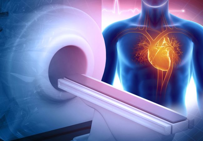
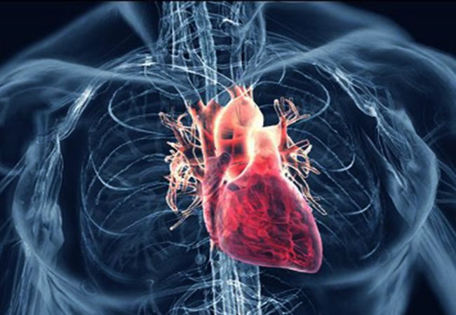
Cardiac MRI
About
Since June 1, 2021, Dikteon MRI Center is offering specialized Cardiac Magnetic Resonance imaging (CMR) services using state-of-the-art equipment to ensure accurate and reliable diagnoses. CMR exams are non-invasive and provide detailed images of the heart and blood vessels, aiding in the diagnosis and monitoring of various heart conditions.


Cardiac MRI
Is one of the most modern diagnostic techniques in cardiology, which has a documented role in both the diagnosis, as well as the treatment of cardiovascular diseases.
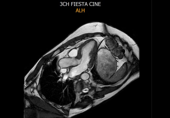
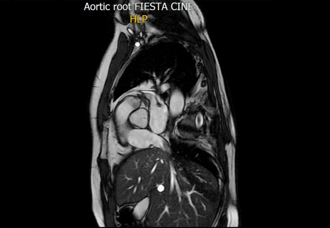
At Dikteon MRI Center, we provide high quality images, with advanced techniques for the detection and evaluation of the entire spectrum of cardiovascular diseases.
The analysis is performed in the most advanced software system CVI42, of the Circle Cadiovascular Imaging Company, allowing for the complete evaluation of each case.
What are the main indications for Cardiac MRI?
Inflammatory diseases of the myocardium. Diagnosis and re-examination.
Cardiomyopathies in storable diseases (Fabry, amyloidosis).
Restrictive cardiomyopathy. Accurate study of myocardial deformity
Evaluation of myocardial vitality and decision to perform coronary angiography
Dilated heart disease to evaluate its functionality of the left and right ventricles (reasons for infarction or damage).
Dilated heart disease to assess functionality (reasons for arrhythmia risk – examination of first degree relative)
Hypertrophic cardiomyopathy. Assessment of arythmia.
Toxic cardiomyopathy. Diagnostis and retesting in patients undergoing or after chemotherapy
Pericardiac diseases. Evaluation of inflammatory disease, detection of cysts or pericardial tumors.
Congenital heart disease in children, adolescents and adults for diagnosis, monitoring and guidance of rehabilitation therapy.
Cardiac Examinations Codes
Cardiac MRI Imaging For Morphology And Function
Cardiac MRI Imaging For Velocity Flow Mapping
Cardiac MRI Imaging Perfusion
Cardiac MRI Imaging For Viability Analysis
